
Macrophage activation syndrome as a complication of dermatomyositis: A case report
2020-06-17 05:54DingXianZhuJianJunQiaoHongFang
World Journal of Clinical Cases 2020年11期
Ding-Xian Zhu, Jian-Jun Qiao, Hong Fang
Ding-Xian Zhu, Jian-Jun Qiao, Hong Fang, Department of Dermatology, The First Affiliated Hospital, College of Medicine, Zhejiang University, Hangzhou 310003, Zhejiang Province,China
Abstract
Key words: Macrophage activation syndrome; Dermatomyositis; Hyperferritinemia; Case report; Systemic juvenile idiopathic arthritis; Inflammatory
INTRODUCTION
Dermatomyositis is an idiopathic inflammatory myopathy characterized by cutaneous and muscular abnormalities. It is a rare and chronic rheumatic disorder, and the reported incidence of dermatomyositis ranged between 2.4 and 13.2 per 100000 population in the United States and Japan[1,2]. Macrophage activation syndrome(MAS), also known as secondary hemophagocytic lymphohistiocytosis, is a potentially life-threatening complication of rheumatic disorders that occurs most commonly in patients with systemic juvenile idiopathic arthritis, systemic lupus erythematosus or adult-onset Still’s disease[3]. The estimated prevalence of MAS in systemic juvenile idiopathic arthritis is around 10%, and the estimated prevalence of MAS among systemic lupus erythematosus patients ranges from 0.9% to 4.6%[4,5].However, few studies have explored MAS as a complication of dermatomyositis. In this report, we describe a patient with dermatomyositis who exhibited fever,hyperferritinemia and central nervous system symptoms, which supported a diagnosis of MAS.
CASE PRESENTATION
Chief complaints
A 44-year-old woman was admitted to our hospital with a 2-wk history of fever with generalized rash, muscle weakness and arthralgia.
History of present illness
The patient’s symptoms started 2 wk ago with a high-grade fever of 39 °C as well as diffuse erythematous and hyperpigmented plaques over the neck, trunk and limbs.
Physical examination
The patient had a limb muscle strength of 2 and reported spontaneous pain in the gastrocnemius muscles.
Laboratory examinations
Laboratory examinations revealed leukocyte count of 21.5 × 10⁹/L, hemoglobin level of 100 g/L, platelet count of 190 × 109/L and negative antinuclear antibody findings.Blood tests are shown in Table 1. The creatine phosphokinase level (137 U/L) was normal. Blood microbiological culture and antinuclear antibody assays also showed negative findings. Tests for hepatitis B, hepatitis C, HIV, cytomegalovirus and Epstein-Barr virus were negative.
Imaging examinations
The chest computed tomography scans showed interstitial lung disease and pulmonary infection. An initial diagnosis of adult-onset Still’s disease was made, andthe patient was treated with meropenem and 200 mg of methylprednisolone.
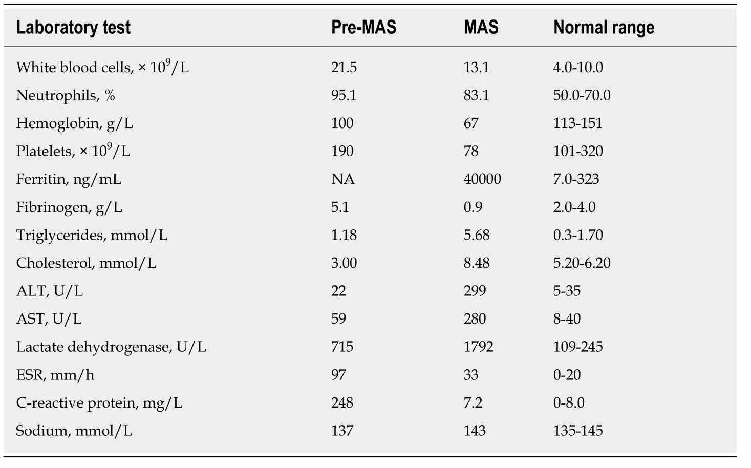
Table 1 Laboratory test results during hospitalization of our patient
Further diagnostic work-up
Fifteen days later, the patient continued to exhibit high fever (38-39.5 °C) and diffuse erythema on the face and neck as well as the Gottron sign over the dorsum of the elbow and knee. Positron emission tomography-computed tomography imaging excluded a diagnosis of tumor but showed an enlarged spleen and swollen axillary lymph nodes. Electromyography findings were suggestive of muscle-derived damage.Although the creatine phosphokinase level was in the normal range, a diagnosis of dermatomyositis was made based on the presence of a typical skin lesion, symptoms of muscle weakness and electromyography findings of muscle-derived damage. The patient had received 200 mg of methylprednisolone for 15 d; however, she continued to exhibit high fever (38-39.5 °C), rash, hepatosplenomegaly, cytopenia, liver dysfunction and coagulopathy. Subsequent hematological examinations showed the following findings: Hemoglobin level, 67 g/L; platelet count, 78 × 109/L; alanine aminotransferase, 299 U/L; aspartate aminotransferase, 280 U/L; ferritin, 40000 ng/mL; and lactate dehydrogenase, 1792 U/L. The fibrinogen level decreased from 5.1 g/L to 0.9 g/L; the triglyceride level gradually increased from 1.18 mmol/L to 5.68 mmol/L, and the cholesterol level increased from 3.0 mmol/L to 8.48 mmol/L (Table 1). Blood cultures remained negative, and the C-reactive protein level normalized.
FINAL DIAGNOSIS
Although the creatine phosphokinase level was in the normal range, a diagnosis of dermatomyositis complicated by MAS was made based on the presence of a typical skin lesion, symptoms of muscle weakness, electromyography findings of musclederived damage, hepatosplenomegaly, cytopenia, liver dysfunction and strikingly high serum ferritin level.
TREATMENT
The patient received 200 mg of methylprednisolone for nearly 20 d.
OUTCOME AND FOLLOW-UP
Unfortunately, the patient’s condition continued to deteriorate, and the patient developed neurological symptoms, including memory loss and convulsions. Head magnetic resonance imaging findings were normal. The patient’s family decided to end treatment, and the patient died.
DISCUSSION
In this report, we have described typical MAS as a complication of dermatomyositis.Our patient presented with persistent fever, high ferritin level and central nervous system symptoms, which are typical clinical features of MAS. To the best of our knowledge, there have been few reports of MAS in patients with dermatomyositis.This report was written to raise awareness of this fatal complication that can develop in patients with dermatomyositis. Importantly, misdiagnosis may delay treatment and result in increased mortality.
MAS has rarely been encountered in patients with inflammatory myopathies; most reported cases in the literature involved patients with juvenile dermatomyositis. In 2019, a systematic review found 12 patients who had both MAS and juvenile dermatomyositis[6]. To our knowledge, there have been only a few cases of MAS developed in adult dermatomyositis reported in English[7,8]. Bazan-Sochaet al[9]reported a fatal case of MAS in a 53-year-old female patient that developed in the early phase of initially mild dermatomyositis with the anti-Jo-1 antibody. Langeet al[10]presented an adult with dermatomyositis who complicated with MAS and the patient died shortly after the confirmation of MAS. It is important to identify MAS as a fatal complication, especially in patients with high serum ferritin levels. Our patient developed central nervous system symptoms. Lillebyet al[11]reported central nervous system involvement in a patient with juvenile dermatomyositis complicated by MAS.Dermatomyositis is rarely associated with the central nervous system but it may be a clinical manifestation of cerebral MAS.
MAS is considered a secondary form of hemophagocytic lymphohistiocytosis; this condition is a rare life-threatening disorder caused by a cytokine storm. It occurs most commonly in patients with systemic juvenile idiopathic arthritis or systemic lupus erythematosus[12]. Clinical features of MAS include fever, cytopenia, hepatosplenomegaly, lymphadenopathy and coagulopathy, followed by multiple organ failure. These symptoms often lead to misdiagnosis of sepsis or disseminated intravascular coagulation[13]. A uniform diagnostic criterion for MAS is not yet available. The most commonly used diagnostic criteria (i.e., “HLH-2004”) include fever, splenomegaly, cytopenia in at least two cell lines, hypertriglyceridemia and/or hyperfibrinogenemia, tissue presentation of hemophagocytosis, minimal or no natural killer-cell activity, serum ferritin concentration > 500 ng/mL and elevated levels of soluble CD25 (> 2 standard deviations above the normal mean; typically > 2400 IU/mL)[14]. Our patient met five of the above diagnostic criteria as well as diagnostic criteria for systemic juvenile idiopathic arthritis[15]. Though hyponatremia is a common clinical manifestation of MAS, no significant difference in the plasmatic sodium was observed in our patient. Notably, our patient exhibited a high serum ferritin level (40000 ng/mL). Serum ferritin is an important laboratory hallmark of MAS and is a well-known predictor of acute exacerbation of interstitial lung disease and poor prognosis in patients with dermatomyositis[16]. Previous studies have shown that activated macrophages contribute to the pathogenesis of acute exacerbation of interstitial lung disease in patients with dermatomyositis[17]. Our findings suggest that macrophage activation may play a role in the pathogenesis of dermatomyositis and that high serum ferritin levels can predict the risk of MAS in patients with dermatomyositis.
The initial rash in our patient was not a typical heliotrope or Gottron rash, and the creatine phosphokinase level was within the normal range. Thus, an initial diagnosis of adult-onset Still’s disease was made. The patient then exhibited typical rash and symptoms of muscle weakness, spontaneous muscle pain and myogenic changes on electromyography. Based on these symptoms, a diagnosis of dermatomyositis was made, in accordance with the criteria of Bohanet al[18]. MAS seemed to occur early in the clinical course of rheumatic disorders. In our case, the occurrence of MAS preceded with the definite diagnosis of dermatomyositis. The clinical manifestations of MAS can overlap with symptoms of dermatomyositis such as rash, fever and arthralgia. However, the changes in hemoglobin, platelet, fibrinogen, ferritin,triglyceride and central nervous system were rarely associated with dermatomyositis,which led to a diagnosis of MAS. Thus, our patient was diagnosed with MAS as a complication of dermatomyositis. Although MAS is considered a rare complication of dermatomyositis, it complicates correctly diagnosing dermatomyositis. Misdiagnosis may delay treatment and result in severe mortality. Literature reported the HLH-2004 criteria did not seem to be sensitive enough, and the clinical suspicion of MAS should be required in the cases with severe hyperferritinemia and its rapid increase[19].
The triggers of MAS in rheumatic disease include two different entities: Cases related to an active infection and cases associated with the onset or flare of the underlying disease[5]. Epstein-Barr virus was the most common causative agent in which the type of infection was reported. Additionally, MAS was reported to occur most frequently in the setting of active systemic juvenile idiopathic arthritis or during a flare of it[4]. In our patient, the Epstein-Barr virus and the blood cultures were negative. We noted that C-reactive protein in our patient decreased from 248 mg/L before the development of MAS to 7.2 mg/L when MAS happened, which was consistent with the literature reporting that low C-reactive protein supports the diagnosis of MAS[20]. The trigger of MAS in our patient was the progressive deterioration of dermatomyositis. Also, our patient showed a poor prognosis after the onset of MAS, suggesting that the MAS was associated with dermatomyositis disease activity.
Based on the findings in this case, MAS should be considered in patients with dermatomyositis. There is a need to increase awareness of this life-threatening condition associated with dermatomyositis, especially in patients who present with a fever and high ferritin levels. However, further studies are needed to explore the prevalence and clinical features of this fatal complication.
CONCLUSION
MAS is an important, potentially fatal, complication of dermatomyositis. Although MAS is rare in dermatomyositis, it should be considered in the differential diagnosis of an unexplained change of hemoglobin, platelet, fibrinogen, ferritin and triglyceride,which may complicate dermatomyositis.
 World Journal of Clinical Cases2020年11期
- World Journal of Clinical Cases的其它文章
- Tumor circulome in the liquid biopsies for digestive tract cancer diagnosis and prognosis
- Isoflavones and inflammatory bowel disease
- Cytapheresis for pyoderma gangrenosum associated with inflammatory bowel disease: A review of current status
- Altered physiology of mesenchymal stem cells in the pathogenesis of adolescent idiopathic scoliosis
- Association between liver targeted antiviral therapy in colorectal cancer and survival benefits: An appraisal
- Peroral endoscopic myotomy for management of gastrointestinal motility disorder