
Comment on: Optic neuropathy and increased retinal glial fibrillary acidic protein due to microbead-induced ocular hypertension in the rabbit
2023-06-17 06:51MengZhenXieYaoZhangYingPingDengLiTang
Meng-Zhen Xie, Yao Zhang, Ying-Ping Deng, Li Tang
1Department of Ophthalmology, West China Hospital, Sichuan University, Chengdu 610041, Sichuan Province, China
2West China School of Medicine, Sichuan University, Chengdu 610041, Sichuan Province, China
Dear Editor,
W e read with great interest the study by Zhaoet al[1],which evaluated optic neuropathy and changes in the amount of glial fibrillary acidic protein in the retina with intracameral injection of microbeads into rabbits.The objective was to investigate whether the injection of microbeads into the anterior chamber can induce chronic intraocular hypertension in a glaucoma model in rabbits.Through dose selection,the researchers finally determined to select 50 μL with a diameter of 15 μm to be injected into the anterior chamber of New Zealand rabbits, and the peak intraocular pressure(23.83±1.14 mm Hg) was reached on day 15.The second peak(23.26±1.03 mm Hg) was reached on day 30 after the initial injection.Intraocular pressure decreased to baseline 60d after the initial injection.They conclude that a simple strategy has been established to induce increased intraocular pressure with associated glaucoma changes in the optic nerve and retina.
We modeled chronic intraocular hypertension in the same way as the researchers did.We injected microbeads (a diameter of 15 μm; FluoSpheres; Ⅰnvitrogen, Carlsbad, CA, USA) with different concentrations, and doses (total injection volume was no less than that of the original author) into the anterior chamber of rabbits (Figure 1), but the intraocular pressure was reduced to baseline within a week.A few days after injection,the number of microbeads in the anterior chamber of rabbits decreased significantly, and only a few could be seen in the iris and the anterior chamber angle.When the rabbit anterior chamber angle was sectioned for many times, no clusters of microbeads were found in the structure (Figure 2).
When we searched literatures, we found that the rabbit had many pectinate ligaments in the filtration angle, large spaces between these ligaments allows aqueous inflow to the trabeculum[2].Its trabeculum contain much less meshes than primates, and rabbit trabecular meshwork was shallow and loose[2-3].The drainage of aqueous filtered through the meshwork is served by a plexus of 1 to 4 channels, which contain giant vacuoles along their internal and external walls[2].In fact, the rabbit eye does not possess the canal of Schlemm but is provided instead with an aqueous plexus which are analogous to the Schlemm in primates[2,4].

Figure 1 The rabbit anterior chamber after injecting microbeads.

Figure 2 Section of rabbit anterior chamber angle 2wk after microbead injection.
In our rabbit study, chronic intraocular hypertension was not maintained, and angular sections did not show microbead aggregation.The aqueous plexus is located outside the trabecula and is formed by 1 to 4 blood vessels in a circular shape, which in some parts circulates with the eye veins.In some places, the walls are very thin and fragile, and the size of the lumen varies widely between about 20 μm and 80 μm[2].And in the rabbit’s eyes, red blood cells can pass through the corner of the chamber[5-6].Combined with the above literature,we speculated that the microbeads might flow into the blood vessels through the space between the chambers.
Other researchers have found that the fibers of trabecular mesh are regular, the surface of collagen fibers is smooth and interwoven into a network, the size of the mesh is different, and pigment particles and red blood cells can be seen in the mesh[3].Even though the researchers didn’t specify the size of the mesh, based on the scale shown in the image, it’s reasonable to assume that the mesh could fit through a microbead with a diameter of 15 μm.
In conclusion, we believe that whether a stable model of chronic ocular hypertension can be created by injecting 15 μm microbeads into the rabbit anterior chamber needs further verification.
ACKNOWLEDGEMENTS
Foundations:Supported by the Cadre Health Research Program of Sichuan Province (No.2023-119); Science &Technology Department of Sichuan Province (China) Funding Project (No.2021YFS0221); 1.3.5 Project for Disciplines of Excellence, West China Hospital, Sichuan University(No.2022HXFH032; No.ZYJC21058; No.2021-023; No.2022-014).
Conflicts of Interest:Xie MZ, None; Zhang Y, None; Deng YP, None; Tang L, None.
Authors Reply to the Editor
Dear Editor,
First, we thank Tanget alfor their interest and attention to our article “Optic neuropathy and increased retinal glial fibrillary acidic protein due to microbead-induced ocular hypertension in the rabbit”.
At the beginning of this study, we went through a challenging exploration in terms of the selection of microbeads injection dosage and technology.From the initial injection volume of 5 μL, an obvious elevation of intraocular pressure was not observed until a single injection of 50 μL microbeads(2.5×10⁵), and the variation of intraocular pressure was shown as Figures 2 and 3 in our paper[1].It’s worth noting that the 50 μL microbeads with the concentration of 2.5×10⁵/50 μL should be injected into the rabbit’s anterior chamber at one time and should not overflow, which is the most important part for the success of the experiment.Tanget almentioned that“total injection volume was no less than that of the original author”in their comment, but they didn’t demonstrate whether the 50 μL microbeads was injected at one time or several times.According to our experience, injection at several times can’t induce the significant elevation of intraocular pressure levels.
Furthermore, we wonder whether Tanget alobserved that the microbeads were especially prone to overflow after intracameral injection.Therefore, enough strategies to prevent the overflow of injected microbeads are required and very important for the success of this experiment.During our experiment, the injection was operated by a doctor skilled in ophthalmic microsurgery.In advance, we performed limbal tunnel puncture under an ophthalmic surgical microscope and discharged most aqueous humor before injecting small bubbles.Then microsyringes were used to inject 50 μL microbeads at one time successfully.The microsyringe should be pulled out quickly and the tunnel mouth area should be pressed gently until there is no leakage.Finally the antibiotic gel was applied inside the conjunctival sac to finish the operation.Tanget aldidn’t describe their detailed operation of intracameral injection and we didn’t know whether it was the same with ours.
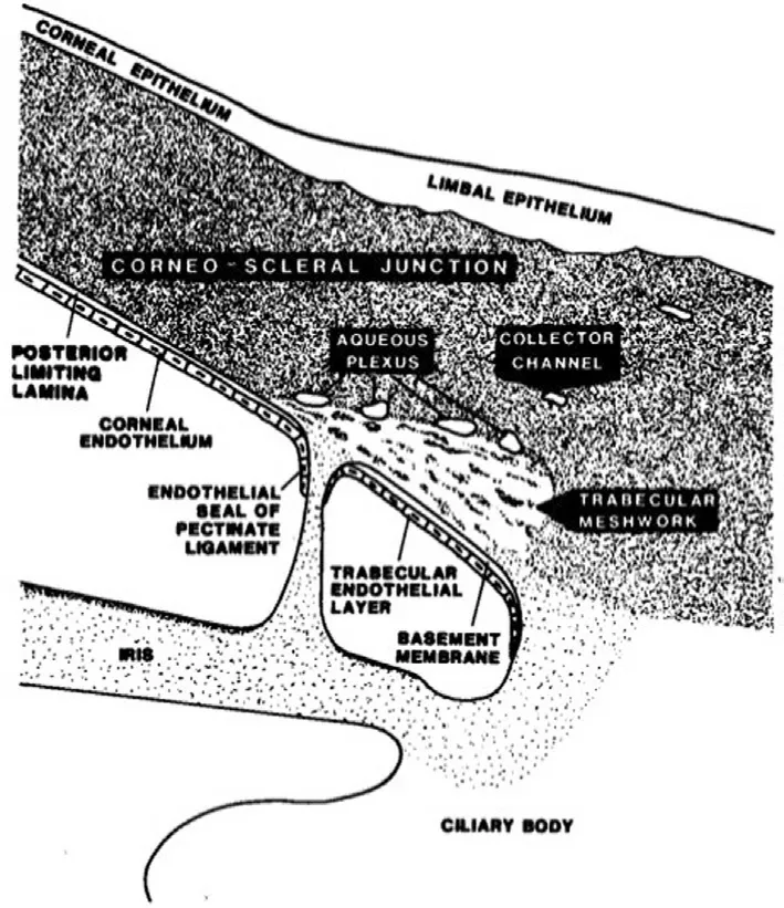
Figure 1 The schematic diagram of rabbit’s anterior chamber angle in the literature[2].

Figure 2 Rabbit’s anterior chamber and microbeads accumulation in the anterior chamber angel[1].
In the appearance, microbeads used in Tanget al’s experiment are light blue, but the microbeads used in our experiment are red and show green under the fluorescence microscope.We wonder whether these two kinds of microbeads have different characteristics, which influence the experimental results.Moreover, it is worthy considering that whether the strain and age of rabbits may also influence the experimental results.In the Figure 2 of Tanget al’s comment, no clear structure of rabbit’s anterior chamber and anterior chamber angle were found, nor was microbeads accumulation.The schematic diagram of rabbit’s anterior chamber angle in the literature was shown in Figure 1[2], which is consistent with our images showing rabbit’s anterior chamber and microbeads accumulation in the anterior chamber angel, as shown in Figure 2[1].
Tian-Hui Zhu1, Jun Zhao1,2
1Shenzhen Eye Hospital Affiliated to Jinan University,Shenzhen Key Laboratory of Ophthalmology, Shenzhen 518040, Guangdong Province, China
2Department of Ophthalmology, Shenzhen People’s Hospital(The Second Clinical Medical College, Jinan University),Shenzhen 518020, Guangdong Province, China
Correspondence to:Jun Zhao.Department of Ophthalmology,Shenzhen People’s Hospital (The Second Clinical Medical College, Jinan University), Shenzhen 518020, Guangdong Province, China.doctorzhaojun@163.com
REFERENCES
1 Zhao J, Zhu TH, Chen WC, Peng SM, Huang XS, Cho KS, Chen DF,Liu GS.Optic neuropathy and increased retinal glial fibrillary acidic protein due to microbead-induced ocular hypertension in the rabbit.Int J Ophthalmol2016;9(12):1732-1739.
2 Bergmanson JP.The anatomy of the rabbit aqueous outflow pathway.Acta Ophthalmol(Copenh) 1985;63(5):493-501.
 International Journal of Ophthalmology2023年6期
- International Journal of Ophthalmology的其它文章
- Preliminary proteomic analysis of human tears in lacrimal adenoid cystic carcinoma and pleomorphic adenoma
- Assessment of the effects of intrastromal injection of adipose-derived stem cells in keratoconus patients
- Evaluation of optic nerve head vessels density changes after phacoemulsification cataract surgery using optical coherence tomography angiography
- Stability of neodymium:YAG laser posterior capsulotomy in eyes with capsular tension rings
- Comparison of the efficacy and safety of ultrasonic cycloplasty vs valve implantation and anti-VEGF for the treatment of fundus disease-related neovascular glaucoma
- Volumetric fluid analysis of fixed monthly anti-VEGF treatment in patients with neovascular age-related macular degeneration