
Effect of acupuncture on acupoint "Yingxiang-Hegu" on Th1, Th2 cytokines and T-bet/GATA-3 of allergic rhinitis rats
2023-12-06 07:54HuQingSuJiaqiLouJinchengMiaoTanyunYinHaoJiMeiqiZhaiChuntaoHaoZhongyaoLuYu
Journal of Hainan Medical College 2023年15期
Hu Qing, Su Jia-qi, Lou Jin-cheng, Miao Tan-yun, Yin Hao, Ji Mei-qi, Zhai Chun-tao, Hao Zhong-yao, Lu Yu-e,?
1.Shanxi University of traditional Chinese Medicine, Jinzhong 030600, China
2.Shanxi Acupuncture Hospital, Taiyuan 030006, China
Keywords:
ABSTRACT Objective: To explore the effect of "Yingxiang-Hegu" on Th1, Th2-related cytokines and [2]transcription factors T-bet and GATA-3 in rats with allergic rhinitis.Methods: Rats were randomly divided into three groups: blank group, model group and acupoint group.The rat model of ovalbumin (OVA) AR was established, and the general condition of the rats was observed and scored.Acupuncture intervention was performed on the acupoint group on the second day after successful modeling, once per day for 20 min for 10 d.After intervention,the general behavior, behavioral score and histomorphological changes of nasal mucosa were observed.Eosinophils (EOS) were counted under microscope after nasal lavage smear staining,and the contents of total IgE, IFN- γ, IL-12, IL-4 and IL-5 in serum were detected by ELISA.Westernblot and IHC were used to detect the protein level and positive protein expression of specific transcription factors T-bet and GATA-3 in rat nasal mucosa.Results: After the establishment of the model, except for the blank group, the behavioral observation scores of rats in the model group and acupoint group were more than 5 points, indicating that the model was successful.After acupuncture intervention on acupoint "Yingxiang-Hegu", the behavioral score of rats in the acupoint group and western medicine group was significantly lower than that in the model group (P<0.05).Microscopic examination showed that the structure of nasal mucosa in the model group was obviously damaged,cilia were arranged discontinuously,uneven, local congestion and swelling, a large number of epithelial cells exfoliated and necrotic, goblet cell proliferation, obvious inflammatory cell infiltration.The pathological degree of nasal mucosa in the pair point group was significantly less than that in the model group.Compared with the model group, the levels of IFN- γ, IL-2 and IL-12 in serum were significantly increased, while IgE, IL-4, IL-5 and IL-6 were significantly decreased, GATA-3 protein and positive expression in nasal mucosa were significantly decreased and T-bet was significantly increased after acupuncture.Conclusion: Acupuncture at "Yingxiang-Hegu" can effectively improve the nasal sensitive symptoms and control nasal inflammation in AR rats.The mechanism may be that acupuncture at Yingxiang-Hegu can up-regulate the expression of T-bet, decrease the level of GATA-3, promote the production of Th1 cytokines and inhibit the synthesis and secretion of Th2 cells, thus restoring the immune balance of Th1 and Th2
1.Introduction
Allergic rhinitis (Allergic Rhinitis,AR), also known as allergic rhinitis, is a non-infectious chronic inflammatory disease of nasal mucosa mediated by immunoglobulin E (IgE) and dominated by type 2 helper T cell (Type2 helper Tcell,Th2) immune response.The main clinical manifestations are paroxysmal sneezing, runny nose, nasal obstruction and itching, and even loss of sense of smell in severe cases[1].The incidence of AR is related to environmental exposure, heredity, climate change, lifestyle and other factors.House dust mite, mold and animal fur are the main sensitization[2-4].In recent years, the prevalence of the disease in China has been on the rise[5].People with AR are prone to occur from children to adults.About 19% to 38% of AR patients are associated with asthma[6].It is also easy to lead to sleep disorders, which brings great pain to patients’ social life, study and work[7].AR is easy to occur repeatedly.The main clinical treatments of AR include drug therapy,immunotherapy, environmental control, health education and so on[8].Acupuncture has certain advantages in the treatment of AR[9].People pay more and more attention to it.At present, it is believed that the main pathogenesis is the immune imbalance between Th1/Th2 and Th17/ regulatory T cells (RegulatoryTcells,Treg)[10].Among them,the imbalance between Th1 and Th2 is considered to be the basis of AR[11].Therefore, the key to the treatment of AR is to adjust the proportion of Th1/Th2 cells and restore the immune balance between Th1/Th2.Professor Lu Jingshan, a master of traditional Chinese medicine, has rich clinical experience in the diagnosis and treatment of AR.Through the application of acupuncture at “Yingxiang-Hegu”,it has achieved remarkable curative effect in the clinical treatment of AR.And the recurrence rate is low, which can effectively improve the quality of life of patients after treatment[12].However, the mechanism of acupuncture on acupoints in the treatment of AR is not clear.The establishment of AR rat model with ovalbumin (Ovalbumin,OVA)can better simulate the clinical symptoms of AR, so this study mainly starts with the immune balance between Th1 and Th2 cells,and observes the effect of acupuncture “Yingxiang-Hegu” on Th1,Th2 cytokines and transcription factor T-bet/GATA-3 in AR rats by constructing OVAAR rat model.To explore the mechanism of acupoint intervention on AR.
2.Materials and methods
2.1 Materials
2.1.1 Experimental animals and groups
Thirty SD rats of 1.1SPF grade, half male and half female, aged 4-6 weeks, (200±40) g,provided by Sibef (Beijing) Biotechnology Co.,Ltd.,[license number:SCXK (Beijing) 2019 Mel 0010].It is raised in the Acupuncture Laboratory of the Scientific Research Building of Shanxi University of traditional Chinese Medicine with a temperature of 20 ℃ and a humidity of 40 ℃ and 60%.After 7 days of adaptive feeding, the rats were numbered from 1 to 30,and the rats were randomly divided into three groups: blank group(n=10),model group (n=10) and point control group (n=10).The rats in the blank group were fed normally without modeling, while the other two groups were made models.The principles of animal ethics were observed during the experiment.This study was approved by the Experimental Animal Ethics Committee of Shanxi University of traditional Chinese Medicine (approval number: AWE202209144).
2.1.2 Main instruments and reagents
Ovalbumin(OVA,A5503, American sigma company);aluminum hydroxide dry powder ([Al(OH)3], A800854, Shanghai Jiyi Biotechnology Co., Ltd.);Rat (IgE) (W20015413),Rat (IL-5)(W20015414) ELISA kit,Huamei biology;IL-4(L220715832),IL-12 (L221010663) ELISA kit, eugenics; Rat IFN-γ (A38020124),Rat IL-2 (A30211236), Rat IL-6 (A30611235) ELISA kit,Novak biology.Actin first antibody, (Actin first antibody, 1HRPgoat anti-rabbit), second antibody HRP- goat anti-rabbit (1RV 5000jjGB23303), Servicebio company; GATA-3 first antibody(1Dre1000 camera AF6233),Affinity company; T-bet first antibody(1Dre1000 camera ab91109), Abcam company.
Enzyme labeling instrument (BioTek, model: Eproch);desktop high-speed freezing centrifuge (Dalong, D3024R);grinder (Servicebio, KZ- II); transfer electrophoresis instrument(Servicebio, SVT-2); ultrasonic cell breaker (Ningbo Xinzhi, JY92-11N);scanner (EPSON, V370); positioned optical microscope(Nikon, NikonEclipseE100); image analysis software (Adobeg,AdobePhotoShop); imaging system (Nikon, NIKONDS-U3, Japan).Disposable aseptic acupuncture needle (specification: 0.25 × 13 mm,Suzhou Medical supplies Factory Co., Ltd.).
2.2 Method
2.2.1 Modeling method
Reference literature[13,14]The modeling method of AR was divided into three stages: 1 basic sensitization: 0.3 mgOVA+30 mgAl(OH)3+1mL saline was made into suspension for backup, each rat in the model group and acupoint group was injected intraperitoneally, and the blank group was replaced by suspension without OVA, once every other day for 7 times in total.2 enhanced excitation: from 14 to 20 d, 50 μL of 5%OVA saline suspension was used for nasal drip on each side, and the blank group was treated with the same amount of saline instead of nasal drip once a day for 7 d.On the 21st to 30th day, 20 μL of 5%OVA sodium chloride solution suspension was given to each side after each intervention 30 min, and the blank group was given the same amount of saline instead of nasal drip once a day for 10 d.(note: because AR is an allergic disease, the body is sensitized after modeling, but the allergy will be triggered only after contact with the allergen, sothe model still needs to be maintained after successful modeling.)
2.2.2 Model evaluation
After the last challenge, the nasal sensitivity symptoms of rats were observed and scored after the last challenge.If the cumulative total score was more than 5, it indicated that the model was successful.
2.2.3 Intervention methods
The intervention began on the second day (that is, the 22nd day)after the end of the model.The blank group was fed normally and did not intervene.Acupuncture intervention was performed on the“Yingxiang-Hegu” acupoints in the acupoint group before each stimulation, and the positioning of Yingxiang and Hegu acupoints referred to the relevant contents of Experimental Acupuncture and moxibustion and Animal Acupoint Map edited by Guo Yi[15].Yingxiang acupoint: when the rats looked directly, the junction of the lateral nostril and the eyeball center, that is, the junction of the lateral nostril and the skin hair of the rat; the Hegu acupoint: located between the first metacarpal bone and the second metacarpal bone of the rat forelimb, when the middle point of the radial margin of the second metacarpal bone.The specific operation is as follows: the rats were tied and fixed after putting on the anti-bite ring, the acupoint area was routinely disinfected, the Hegu acupoint was punctured directly into the needle, and the Yingxiang acupoint was inserted obliquely into the needle, the depth was about 0.3 cm, the needle was retained 20 min, and the Hegu acupoint was synchronously needled,once a day, once a day for 10 d.The model group was the same as the point group without acupuncture.
2.2.4 Behavioral observation score
The symptom superposition quantitative score method was used before and after the establishment of the model, after the last intervention and after nasal stimulation[16],the allergic symptoms of the nose of rats were observed and scored, and the scoring criteria were detailed in Table 1.

Tab 1 scoring criteria for nasal allergic symptoms
2.2.5 Drawing materials
Samples were collected on the second day after the end of the intervention.After weighing and intraperitoneal injection of 3%30 mg/Kg pentobarbital sodium, the blood was collected and centrifuged by 15min (3 000 r/min).The supernatant was removed to the cryopreservation tube.Then the nasal cavity was irrigated with saline through the posterior nasal meatus with a syringe, and the lavage fluid was collected at the nostril with a 1.5 mL EP tube.Then the bilateral nasal mucosa of the rats were stripped, one side was fixed in 4% neutral paraformaldehyde solution, and the other side was frozen with liquid nitrogen wrapped in tin foil and transferred to the refrigerator at-80 ℃.
2.2.6 Histomorphological observation of nasal mucosa
After dehydration, embedding, drying, xylene transparency,dewaxing and washing, the fixed nasal mucosa was stained with HE and sealed with neutral gum, and the histomorphological and pathological changes were observed under microscope.
2.2.7 EOS count of nasal lavage smears
The nasal lavage fluid was centrifuged at 3 000 rpm for 10 min,and the sediment smear was dried and fixed.After paraffin marking,Wright’s dye was added, 1min was placed static, PBS buffer was added, 5min was washed with running water, and EOS count was performed after dry X-ray examination.
2.2.8 The levels of serum total IgE, IFN-γ, IL-2, IL-12,IL-4, IL-5 and IL-6 in rats were detected by ELISA
The collected blood samples were centrifuged at 3 000 r for 10 min,then the supernatant was taken and put in the refrigerator at 4 ℃ to be tested.Then the contents of IgE, IFN-γ, IL-2, IL-12, IL-4, IL-5 and IL-6 in serum of rats were detected according to the instructions of the kit.
2.2.9 Detection of T-bet and GATA-3 protein levels in rat nasal mucosa by Western blot
Extract protein from nasal mucosa by adding lysate homogenate,prepare SDS-PAGE sample electrophoresis, electrophoresis to the bottom of bromophenol blue about 1cm, 300 mA constant current transfer membrane 30 min (wet transfer), wash after transfer and add skim milk to decolorize, seal at room temperature and add primary antibody (GATA-31:1 000;T-bet1:1 000).Actin1:3 000), overnight at 4 ℃, wash the membrane and add the second antibody (HRPgoat anti-rabbit 1vir 5 000).After incubation at room temperature,wash the membrane again, develop the ECL solution, scan the film for archival, collect pictures, and analyze the protein bands of each group.
2.2.10 Immunohistochemical detection of T-bet and GATA-3 expression in rat nasal mucosa
The paraffin sections of nasal mucosa were dewaxed to water in turn, washed on the decolorizing shaker after natural cooling, dripped 3%BSA in the histochemical circle, sealed at room temperature, and incubated at 4 ℃.The slices were incubated at 4 ℃ overnight; the second antibody was added and PBS was washed 3 times at room temperature; the positive area was brownish yellow after dripping DAB chromogenic solution, and the staining was terminated by rinsing with tap water.After re-dyeing, the differentiation solution differentiated, returned to blue, rinsed with running water,dehydrated and transparent, and sealed with neutral gum.The results were interpreted under a white light microscope.The nucleus stained with hematoxylin was blue, and the positive expression of DAB was brownish yellow.
2.2.11 Statistical Analysis
Statistical software SPSS26.0 was used to analyze and process the experimental data.The average absorbance of immunohistochemical results of each group was analyzed by ImageProPlus7.0.Statistical graphs of experimental data were processed by GraphPadPrism8.0 software.All data were expressed as mean ± standard deviation (±s).After normal and homogeneity test of variance, One-WayANOVA was used for mean comparison and LSD-t test was used for comparison between groups.When P < 0.05, the difference was considered statistically significant.
3.Results
3.1 Comparison of body mass of rats before and after modeling and intervention
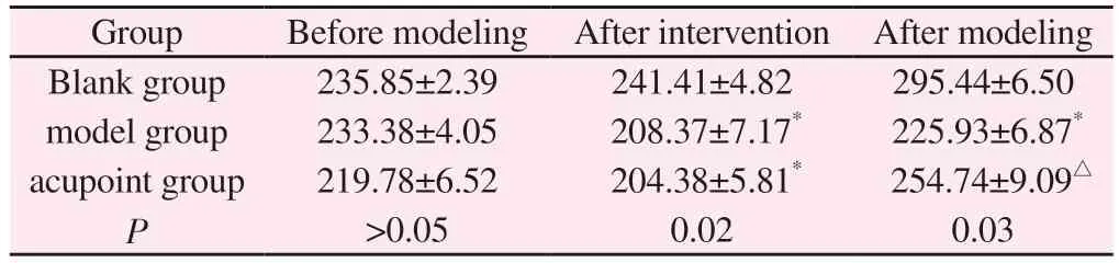
See Table 2.Before modeling, there was no significant difference in body weight among the three groups (P>0.05), but after modeling,compared with the blank group, the body weight in each group decreased significantly (P<0.05), and after acupuncture intervention,compared with the same period in the model group, the body weight of rats in the control group increased significantly (P<0.05).
Tab 2 Comparison of body mass of rats in each group before and after modeling and after intervention(±s, n=10, g)
Tab 2 Comparison of body mass of rats in each group before and after modeling and after intervention(±s, n=10, g)
Note:compared with the blank group in the same period,*P<0.05,compared with the model group,△P<0.05.
Group Before modeling After intervention After modeling Blank group 235.85±2.39 241.41±4.82 295.44±6.50 model group 233.38±4.05 208.37±7.17* 225.93±6.87*acupoint group 219.78±6.52 204.38±5.81* 254.74±9.09△P>0.05 0.02 0.03
3.2 Comparison of nasal symptom scores in rats
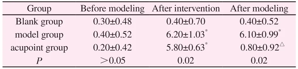
See Table 3.After modeling, except the blank group, all the rats in the model group had nasal allergy symptoms, and the total score of the model group was more than 5, indicating that the model was successful, and the total score of AR symptom behavior in the acupoint group was significantly lower than that in the model group after the last intervention, and there was no significant difference between the acupoint group and the blank group.
Tab 3 Comparison of nasal symptom scores in each group(±s, n=10)
Tab 3 Comparison of nasal symptom scores in each group(±s, n=10)
Note:compared with the blank group in the same period,*P<0.05,compared with the model group,△P<0.05.
Group Before modeling After intervention After modeling Blank group 0.30±0.48 0.40±0.70 0.40±0.52 model group 0.40±0.52 6.20±1.03* 6.10±0.99*acupoint group 0.20±0.42 5.80±0.63* 0.80±0.92△P>0.05 0.02 0.02
3.3 Histomorphological observation of nasal mucosa
See figure 1.Microscopic examination showed that the structure of nasal mucosa in the blank group was intact, cilia were dense, arranged neatly, and there was no obvious infiltration of inflammatory cells, while in the model group, the structure of nasal mucosa was obviously damaged, the arrangement of cilia was discontinuous, uneven, local congestion and swelling, a large number of epithelial cells exfoliated and necrotic, goblet cell proliferation,obvious inflammatory cell infiltration.The pathological degree of nasal mucosa in the point group was significantly less than that in the model group, the structure was relatively complete, no obvious hyperemia and edema, neat cilia, a small amount of loss, orderly arrangement and uniform distribution of epithelial cells, no obvious goblet cell proliferation and a small amount of inflammatory cell infiltration were found in the point group.
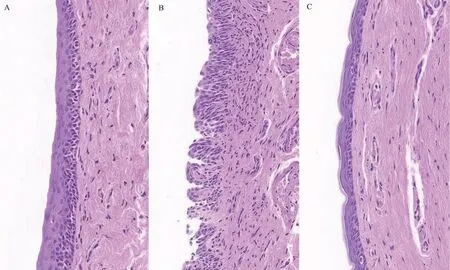
Fig 1 HE staining results of nasal mucosa of rats in each group.
3.4 EOS count of cell smears in nasal lavage fluid
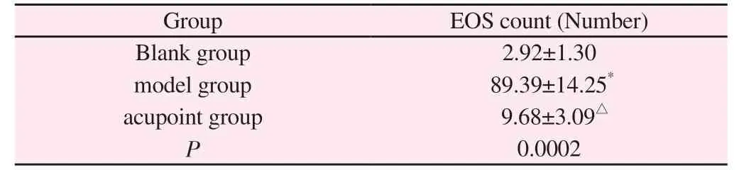
See Table 4.Compared with the blank group, the number of EOS in the model group was significantly increased, but there was no significant difference between the acupoint group and the model group, but the number of EOS in the acupoint group was significantly lower than that in the model group.
Tab 4 Comparison of EOS count in nasal lavage smear of rats in each group(±s,n=10)
Tab 4 Comparison of EOS count in nasal lavage smear of rats in each group(±s,n=10)
Note: compared with the blank group in the same period,*P<0.05,compared with the model group,△P<0.05.
Group EOS count (Number)Blank group 2.92±1.30 model group 89.39±14.25*acupoint group 9.68±3.09△P 0.0002
3.5 Comparison of serum IgE, IFN- γ, IL-2, IL-12, IL-4,IL-5 and IL-6 contents
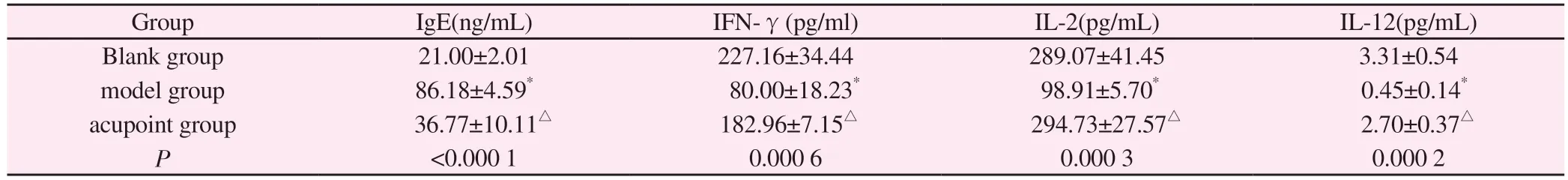
See tables 5 and 6.Compared with the blank group, the contents of IgE, IL-4, IL-5 and IL-6 in the serum of the model group increased significantly, while the levels of IFN- γ, IL-2 and IL-12 decreased significantly.Compared with the model group, the contents of IFN- γ, IL-2 and IL-12 in the serum of rats in the control group increased significantly, while the levels of IgE, IL-4, IL-5 and IL-6 decreased significantly.Compared with the blank group, there was no significant difference in each index between the point group and the blank group (P> 0.05).
3.6 Expression of T-bet and GATA-3 protein specific transcription factors in rat nasal mucosa
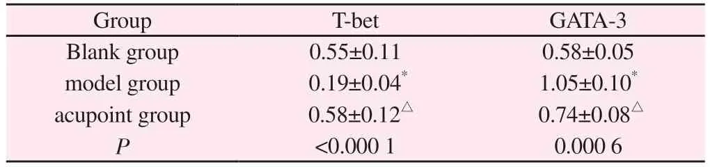
See Figure 2.Compared with the blank group, the GATA-3 protein level in the nasal mucosa of the model group rats was significantly increased, while the T-bet level was significantly reduced (P<0.05);Compared with the model group, the expression of T-bet protein in the acupoint group rats was significantly increased, while the level of GATA-3 was significantly reduced (P<0.05).Compared with the blank group, there was no significant difference in the expression of T-bet and GATA-3 proteins in the acupoint group (P>0.05).
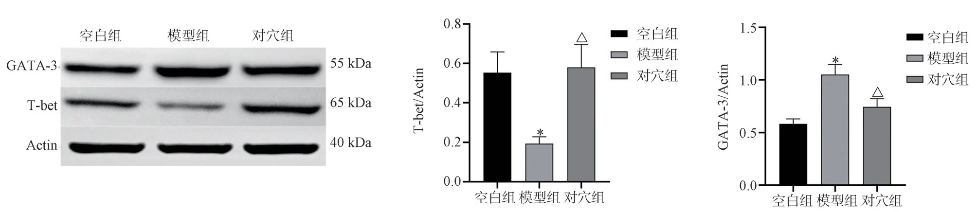
Fig 2 Comparison of T-bet and GATA-3 protein expression in the nasal mucosa of rats in each group(±s, n=10)
Tab 5 Comparison of serum IgE, IFN- γ, IL-2 and IL-12 contents in rats (±s, n=10)
Tab 5 Comparison of serum IgE, IFN- γ, IL-2 and IL-12 contents in rats (±s, n=10)
Note:compared with the blank group in the same period,*P<0.05,compared with the model group,△P<0.05.the same below.
Group IgE(ng/mL) IFN-γ(pg/ml) IL-2(pg/mL) IL-12(pg/mL)Blank group 21.00±2.01 227.16±34.44 289.07±41.45 3.31±0.54 model group 86.18±4.59* 80.00±18.23* 98.91±5.70* 0.45±0.14*acupoint group 36.77±10.11△ 182.96±7.15△ 294.73±27.57△ 2.70±0.37△P<0.000 1 0.000 6 0.000 3 0.000 2
3.7 Immunohistochemical staining method for detecting the positive expression of T-bet and GATA-3 in rat nasal mucosa
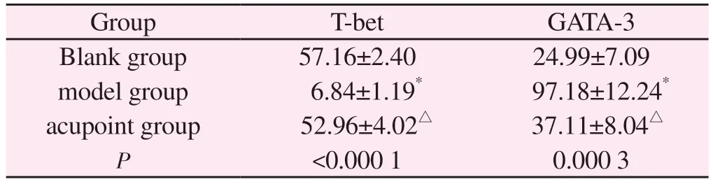
See Figures 3 and 4.The brownish yellow part is the positive expression area.The brownish yellow part is the positive expression area From the positive expression of T-bet, there were brown areas in the cytoplasm of inflammatory cells in the lamina propria of the nasal mucosa in the blank group, suggesting that the expression of T-bet in normal nasal mucosa was high; The brown yellow staining range of the model group is relatively small, and the expression level of T-bet is relatively low; The range of positive expression in the acupoint group is relatively large, and the level of T-bet expression is higher than that in the model group From the positive expression of GATA-3, GATA-3 is more expressed in the cytoplasm.In the blank group, nasal mucosa tissue has a small range of brown yellow areas in the lamina propria, and the staining is light, which indicates that GATA-3 is less expressed in normal nasal mucosa tissue; In the model group, there was a large area of positive areas in the cytoplasm of inflammatory cells in the lamina propria of the nasal mucosa, which were dark brown in color, and the expression of GATA-3 was high; In the contra acupoint group, a small area of brownish yellow area can be seen in the lamina propria of the rat na-sal mucosa, the color is lighter, and GATA-3 is low expressed.③From the average optical density, the average absorbance of GATA-3 in the model group was significantly increased compared to the blank group, while T-bet was significantly decreased (P<0.05).There was no significant difference in the positive expression of T-bet and GATA-3 in the acupoint group (P>0.05); Compared with the model group, the average absorbance of GATA-3 in the acupoint group was significantly reduced, and the expression of T-bet was significantly increased (P<0.05).
Tab 6 Comparison of IL-4, IL-5, and IL-6 levels in rat serum(±s, n=10)

Tab 6 Comparison of IL-4, IL-5, and IL-6 levels in rat serum(±s, n=10)
Group IL-4(pg/mL) IL-5(pg/mL) IL-6(pg/mL)Blank group 63.07±2.84 24.99±7.09 61.17±11.01 model group 134.25±24.90* 97.18±12.24* 161.35±21.70*acupoint group 83.99±6.92△ 37.11±8.04△ 83.69±7.30△P 0.003 0 0.000 2 0.000 4
Tab 7 Comparison of T-bet and GATA-3 protein expression in rat nasal mucosa(Tbet/Actin,GATA-3/Actin, ±s)
Tab 7 Comparison of T-bet and GATA-3 protein expression in rat nasal mucosa(Tbet/Actin,GATA-3/Actin, ±s)
Note:compared with the blank group in the same period,*P<0.05,compared with the model group,△P<0.05.
Group T-bet GATA-3 Blank group 0.55±0.11 0.58±0.05 model group 0.19±0.04* 1.05±0.10*acupoint group 0.58±0.12△ 0.74±0.08△P<0.000 1 0.000 6
Tab 8 Comparison of average absorbance of T-bet and GATA-3 in rats of each group(±s,n=10)
Tab 8 Comparison of average absorbance of T-bet and GATA-3 in rats of each group(±s,n=10)
Group T-bet GATA-3 Blank group 57.16±2.40 24.99±7.09 model group 6.84±1.19* 97.18±12.24*acupoint group 52.96±4.02△ 37.11±8.04△P<0.000 1 0.000 3
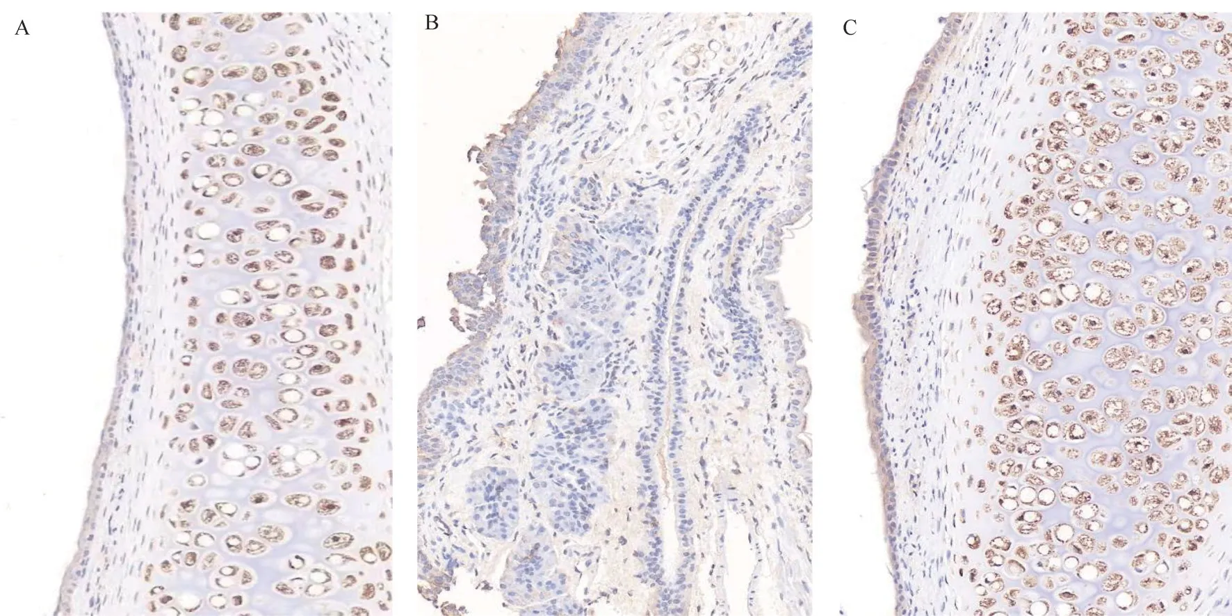
Fig 3 Comparison of T-bet positive expression results in nasal mucosa tissues of rats in each group
Fig 4 Comparison of GATA-3 positive expression results in nasal mucosa tissues of rats in each group
4.Discussion
Allergic rhinitis (AllergicRhinitis,AR) is also called allergic rhinitis.According to its clinical manifestations, traditional Chinese medicine classifies it into the category of “nasal sneeze” and “sneeze”.The name of the disease “rhinorrhea” comes from the explanation of Su Wen’s pulse: “the so-called guest and grandson pulse.” Yangming is at the same time, so it has headache, nasal pain and abdominal swelling[17].”In the aspect of acupoint selection, acupuncture and moxibustion is mainly used in the treatment of allergic rhinitis by local acupoint selection and distal acupoint selection, while in meridian selection, acupoints on Yang meridian are mainly selected,and the diversification of acupoints is advocated[18~22].Under the influence of the theory of “medicine to medicine” by Professor Lu Jingshan, a master of Chinese medicine, Professor Lu Jingshan,a famous doctor in Beijing, has systematically concluded that it has a wide clinical basis after years of clinical practice.Lu Lao has achieved remarkable curative effect in the treatment of AR by acupuncture “Yingxiang-Hegu”, which is widely praised by patients[21].Yingxiang acupoint is an important point for acupuncture and moxibustion in the treatment of rhinopathy.The first and second Classic of Acupuncture and moxibustion says: “the nose is unfavorable, asphyxiated, remote, runny, bleeding has carbuncle,welcome fragrance.” Yingxiang acupoint is located beside the nose and belongs to local acupoint selection.Under the acupoint is the distribution of anastomotic branches of facial artery and vein and facial nerve and infraorbital nerve.Needling can promote the nasal orifices, restore the sense of smell, and Yingxiang belongs to the hand and foot Yangming meridian intersection points, so it can regulate the qi of the two meridians, dispel the wind and clear the orifices, improve the nasal microcirculation, make the nasal channels unobstructed, and dredge the qi of the exterior and interior meridians-the lung meridians, enhance the descending power of the lungs, and the lungs pass through the nose, so it can clear the nose orifices.The research shows that acupuncture[22],acupoint injection[23,24], acupoint application[25], buried wire[26] at Yingxiang point have achieved satisfactory results in the treatment of AR,the reason may be related to the fact that acupuncture at Yingxiang point can restore the clearance function of ciliated layer in nasal mucosa.Hegu acupoint is located in the back of the hand, at the radial midpoint of the second metacarpal bone.It is the original acupoint of the large intestine meridian of hand Yangming.It has the function of opening and purging Yangming, soothing the wind and relieving the surface, clearing and purging lung qi, and treating a variety of diseases, mainly exogenous pathogens, head facial features and other diseases.“Mian Kou Hegu Shou” in “Acupuncture and moxibustion Dacheng” points out that Hegu acupoint can treat many facial and oral diseases.The large intestine meridian of the hand Yangming and the lung meridian of the hand Taiyin are on the surface, the lung dominates the fur, and the external sensation shows all the diseases.Yingxiang is mainly to dredge the qi of the nasal meridian, and Hegu focuses on purging the qi of the meridians.The two acupoints match each other[27].The research results of Li Lingxin[28] et al show that there is a correlation between the change of body surface infrared thermal image temperature after acupuncture at Hegu acupoint and the body surface of Yingxiang acupoint on the opposite side,suggesting that the meridian route is specific.The research of Zheng Meifeng[29] and others shows that acupuncture Yingxiang and Hegu can regulate the unbalanced neurotransmitter function and relieve the symptoms of AR, but the specific mechanism of the combination of Yingxiang and Hegu in the treatment of AR is not clear.
Studies have shown that the immune imbalance of Th1/Th2 caused by the dominance of Th2 immune response is considered to be the basis of the pathogenesis of AR[30], while the imbalance of Thl/Th2 proportion is shown by the changes of related cytokines, such as IL-12, IFN-γ, IL-4, IL-5 and so on.Therefore, inhibiting the production of Th2 cytokines and regulating the balance of Th1, Th2-related cytokines and transcription factors may be the key to the treatment of AR[31].IgE can degranulate mast cells and BAS, release a variety of inflammatory mediators, aggravate the inflammatory response, and play an important role in the pathogenesis of AR[32].As Th1 cytokines, IFN-γ, IL-2 and IL-12 can not only reduce the immune activity of BAS, but also reduce the production of IgE and inhibit the degranulation of mast cells, so as to inhibit the immune drive of Th2 and promote the secretion of Th1 cells,so that the immune balance of Th1/Th2 can be further regulated,and then slow down the process of AR[33~38].IL-4, IL-5 and IL-6 are Th2 cytokines.By promoting the synthesis and secretion of IgE, stimulating mast cells and enhancing the proliferation and infiltration of EOS, they mainly play a pro-inflammatory role in Th1/Th2 immune balance, inhibit the expression of Th1 cytokines IL-2, IL-12 and IFN-γ,and prevent them from participating in the process of Th0 cells differentiating into Th1 cells, thus enhancing the occurrence and progress of inflammatory response characterized by Th2[39,40].Th2-specific transcription factor GATA-3 mainly affects the transcription of Th2 cytokines, which not only promotes the secretion of IL-4 and inhibits the production of IFN-γ, but also prevents primordial T cells from differentiating into Th1 cells,making the body in an allergic inflammatory state dominated by Th2 cell immune response, resulting in the clinical symptoms of AR[41].T-bet is a specific transcription factor for Th1 cell differentiation,which can promote the production of IFN-γ, induce Th2 cells to differentiate into Th1 cells, reduce the proportion of Th2 cytokines,and thus inhibit inflammation[42].
The results of this experimental study showed that the scores of nasal symptoms of rats increased significantly after modeling, and all of them were more than 5 points, indicating that the model was successful, and the levels of Th1-related cytokines IFN-γ, IL-2 and IL-12 in serum of rats in the model group were significantly decreased, while the levels of Th2 cytokines IL-4, IL-5 and IL-6 were significantly increased, which further verified that the immune imbalance of Th1/Th2 was involved in the pathogenesis of AR, which was consistent with the previous research results[43].After acupuncture intervention on acupoint “Yingxiang-Hegu”,the behavioral score of AR rats in the acupoint group decreased significantly, and the EOS count in nasal lavage fluid decreased significantly, the inflammatory infiltration of nasal mucosa was significantly improved, the expression of GATA-3 in nasal mucosa was significantly decreased, the level of T-bet was significantly decreased, and the levels of IFN-γ, IL-2 and IL-12 in Th1 cells were significantly increased.The levels of Th2 cytokines IL-4, IL-5 and IL-6 decreased significantly, which corrected the immune imbalance between Th1/Th2 cells and alleviated the local inflammatory response.Therefore, the intervention effect of acupuncture on acupoint “Yingxiang-Hegu” may be by reducing the level of Th2-specific transcription factor GATA-3, preventing Th0 cells from differentiating into Th2 cells, and then reducing the secretion of Th2 type cytokine pro-inflammatory factors IL-4, IL-5 and IL-6.At the same time, it can increase the expression of Th1-specific transcription factor T-bet, promote the differentiation of Th1 cells and play an immunosuppressive role, so that Th1/Th2 can restore immune balance.This study mainly discusses the effect of acupuncture on acupoint “Yingxiang-Hegu” on Th1/Th2-related cytokines and specific transcription factors in AR rats, expounds the mechanism of acupuncture on acupoints interfering with AR, and provides some evidence of evidence-based medicine for clinic.
 Journal of Hainan Medical College2023年15期
- Journal of Hainan Medical College的其它文章
- Establishment of extensively drug-resistant Pseudomonas aeruginosa pneumonia model in rat
- MiR-873 regulates cell autophagy by targeting Beclin1 to promot inflammation and apoptosis of bronchial epithelial cells
- Monitoring and analysis of contamination of Vibrio parahaemolyticus and Vibrio alginolyticus in seafood in Haikou
- Research progress on cardiotoxicity mechanism of doxorubicin and prevention and treatment of traditional Chinese medicine
- Study on regulating mechanisms of oxocrebanine obtained from Stephania hainanensis H.S.Lo et Y.Tsoong on microtubule sites and tubulin in human breast cancer MCF-7 cells
- Study on the mechanism of Fuzi in the treatment of allergic rhinitis based on network pharmacology and experimental validation